Tantalum (Ta) is a rare transition metal element discovered by Swedish chemist Anders Gustav Ekeborg in 1802, with atomic number 73 [1]. Tantalum-based materials refer to a class of materials primarily composed of tantalum or utilizing it as a core structural unit. These materials are modified through alloying, compositing, or specialized processing techniques to meet specific application requirements. They encompass tantalum, porous tantalum, tantalum alloys, tantalum compounds, and tantalum-based composites. In recent years, tantalum-based materials have played an irreplaceable role in critical fields such as chemical corrosion protection, electronic capacitors, aerospace, and military applications due to their unique physicochemical properties, exceptional corrosion resistance, outstanding high-temperature stability, and excellent thermal and electrical conductivity. Simultaneously, their superior biocompatibility and mechanical adaptability have revealed new application potential in the biomedical field. With further breakthroughs in material modification technologies, tantalum-based materials have rapidly expanded their applications in orthopedics, dentistry, imaging, and cardiovascular interventional procedures, becoming one of the core materials for high-end implant devices. This paper systematically reviews the biological basis, current status, and future directions of tantalum-based materials in medical applications, providing a reference for their widespread clinical utilization.
1 Biological Basis for Tantalum-Based Materials in Medical Applications
Tantalum-based materials exhibit exceptional biological properties, including excellent osseointegration, hemocompatibility, and antimicrobial performance. These characteristics are intrinsically linked to their unique material properties and form the biological foundation for their extensive medical applications.
1.1 Osseointegration Properties of Tantalum-Based Materials
Osseointegration refers to the direct connection formed between an implant and bone tissue, involving multiple biological processes. Upon implantation into bone, the device triggers a localized inflammatory response. Subsequently, an osteoid layer forms on the implant surface, inducing bone tissue proliferation and differentiation. This leads to a tight bond between the two, ultimately achieving stable integration between the bone and the implant. This process is intrinsically linked to both the implant material itself and its surrounding environment. Tantalum is a biologically inert metal that does not stimulate bone growth. However, its derivatives—such as tantalum oxide, porous tantalum, and nanotantalum—exhibit exceptional osseointegration properties. These materials promote osteoblast proliferation and differentiation while facilitating inward growth of bone, tendons, and ligaments.
In cytological studies, researchers prepared Ta₂O₅ coatings on titanium implant surfaces. Following specialized treatment, these Ta₂O₅ coatings enhanced osteoblast and fibroblast adhesion, proliferation, differentiation, mineralization, and expression of osteogenic genes [2-3]. Co-culturing porous tantalum with bone marrow mesenchymal stem cells, MG63 osteoblasts, and dental pulp stem cells to evaluate its bioactivity revealed that porous tantalum significantly enhanced cell adhesion, proliferation, and osteogenic differentiation [4]. Other studies similarly demonstrated tantalum's non-toxicity toward L929 mammalian cells, human monocytic leukemia cells, gingival fibroblasts, and fibroblasts [5-8], while enhancing peri-implant soft tissue integration. One study reported that tantalum-coated implants immersed in phosphate-buffered saline for 28 days released 0.2 μg/L of tantalum. However, in clinical applications, mechanical wear inevitably causes nanoscale debris to detach from the implant surface, potentially affecting the survival of surrounding cells. Regarding these nanoscale tantalum particles, Wang et al.[9] found that low concentrations (12.5 μg/mL) of tantalum nanoparticles promote osteoblast proliferation, whereas concentrations ≥25 μg/mL begin to reduce osteoblast activity. This aligns with recent studies [10-12], collectively confirming tantalum nanoparticles exhibit no cytotoxicity and significantly promote osteoblast adhesion, proliferation, maturation, and differentiation at optimal concentrations. Recent studies have revealed that tantalum interacts with numerous classical osteogenic signaling pathways, including the Wnt/β-catenin pathway, transforming growth factor-β and osteomorphin pathways, mitogen-activated protein kinase pathway, and integrin signaling pathway [13]. Compared to titanium, tantalum induces higher expression of key molecules in osteogenic signaling pathways, indicating tantalum exerts more potent effects on multiple osteogenesis-related signaling pathways. These studies provide crucial mechanistic insights into tantalum-induced bone formation.
In animal experiments, Wei et al. [14] implanted porous tantalum rods into the hind legs of dogs. After 3 to 6 weeks, biopsy observations revealed the adhesion of new osteoblasts and the ingrowth of new bone at the tantalum-host bone interface and within the pores. Additionally, Chinese researchers implanted porous tantalum and porous titanium implants into bilateral femoral condyle defects created in New Zealand White rabbits. Observations at 2, 4, and 8 weeks revealed that porous tantalum formed early biological bonding with bone tissue, demonstrating osteointegration capabilities comparable to porous titanium [15]. Similarly, Fraser et al. [16] observed that although porous tantalum implants did not significantly affect implant stability coefficients, hardness, or the elastic modulus of newly formed bone in rabbits, tantalum-based implants demonstrated significantly higher levels of neovascularization, wound healing, and expression of osteogenesis-specific genes compared to titanium-based implants [17]. In a recent report, Zhang et al. [18] implanted porous tantalum fusion devices—produced via 3D printing and chemical vapor deposition—into the C3/4 cervical spine segments of sheep. After 12 months of follow-up, both groups demonstrated excellent osteogenic outcomes and long-term biocompatibility. This study not only demonstrates the osseointegration properties of tantalum-based materials but also suggests that 3D-printed tantalum-based materials can achieve complex structural designs, low-cost manufacturing, and personalized customization, providing a solid scientific basis for the broader clinical application of tantalum-based materials in the future.
1.2 Hemocompatibility of Tantalum-Based Materials
Tantalum-based materials exhibit significant advantages in hemocompatibility, particularly in stents and other blood-contacting implants. Their superior properties—including enhanced endothelialization, thrombosis inhibition, low platelet adhesion and activation, and prolonged clotting time—make them ideal materials for blood-contacting applications. Tantalum's surface charge, corrosion resistance, and semiconductor properties play a crucial role in preventing thrombosis and promoting vascular healing. These characteristics provide strong support for tantalum applications in cardiovascular, orthopedic, and other fields.
Endothelialization refers to the process where vascular endothelial cells form a continuous cell layer on the stent surface. This process inhibits platelet aggregation by releasing anticoagulant substances such as prostacyclin and nitric oxide, which is crucial for preventing thrombus formation and maintaining normal vascular function. It is a key factor in ensuring the long-term safety of arterial stents. Giessen et al. [19] implanted a sinusoidal coil stent made from 0.127 mm diameter tantalum wire into animal arteries. This stent achieved full endothelialization within 7 days, effectively preventing thrombus formation. In another study, this research group compared the efficacy of self-expanding cobalt alloy stainless steel wall stents versus balloon-expandable tantalum stents in a porcine model. Results showed that the stainless steel stents had a patency rate of 62% at one week, rising to 100% with anti-thrombotic medication. while tantalum stents demonstrated 100% patency at one week without antithrombotic drugs [20]. Chopra et al. [21] cultured periodontal ligament stem cells and endothelial progenitor cells separately or co-cultured on porous tantalum (Ta) metal discs. They found that Ta discs significantly promoted proliferation of both cell types, enhanced osteogenic and neovascular activities, and analyzed that this was achieved by increasing Runt-related transcription factor 2 and vascular endothelial growth factor receptor 2 activity. These studies demonstrate that tantalum scaffolds exhibit superior endothelialization properties, favoring long-term safety in maintaining vascular patency, and show significant advantages in hemocompatibility.
Given tantalum's excellent hemocompatibility, it has also been incorporated into other polymeric, metallic, and ceramic biomaterials to enhance hemocompatibility. Piquet et al. [22] co-wove 0.1 mm tantalum wire with Dacron fibers to fabricate a stent graft for treating aortic aneurysms in minipigs. Six weeks post-implantation, the entire lumen of the stent graft was fully endothelialized, and all grafts remained patent until retrieval, effectively treating the aneurysms. Bakri et al. [23] incorporated tantalum ions onto expanded polytetrafluoroethylene (ePTFE) surfaces via plasma ion deposition. The resulting nanoscale tantalum-enriched surface layer promoted endothelial cell adhesion, proliferation, and spreading while inhibiting platelet adhesion and activation, demonstrating significant advantages over unmodified ePTFE. Furthermore, in a canine aortic bypass model, tantalum-modified ePTFE demonstrated anti-thrombotic properties by significantly inhibiting thrombosis through rapid formation of an intraluminal endothelial cell layer.
1.3 Antibacterial Properties of Tantalum-Based Materials
The antimicrobial properties of materials can effectively reduce the incidence of related infections, thereby improving the success rate of implant surgeries. Ionic tantalum exhibits satisfactory antibacterial capabilities, with its mechanism potentially involving the combined or separate effects of enzyme disruption, DNA denaturation, cell wall/cell membrane degradation, and signaling pathway disruption. In a recent study, Singh et al. [24] surface-modified dental implant materials (CPTi Grade 4 and Grade 5 titanium alloys) with tantalum coatings. They found this modification significantly reduced the growth of Staphylococcus aureus and Candida albicans, demonstrating that tantalum-coated modifications substantially enhance the antimicrobial properties of implants. However, porous tantalum did not exhibit anti-biofilm properties in certain in vitro experiments. Therefore, the antibacterial properties of tantalum remain inconclusive.
To improve the clinical application of tantalum-based materials, researchers employ surface modification techniques to enhance the antibacterial performance of tantalum coatings or tantalum-based implants. Wang Cuicui et al. [25] doped Cu into Ta₂O₅ nanorods on the tantalum surface and investigated their antibacterial properties, finding that Cu doping significantly enhanced the antibacterial activity of porous tantalum. Ding et al. [26] utilized magnetron sputtering technology to prepare a multilayer composite coating of ZnO-doped tantalum oxide at room temperature. comprising a five-layer structure from inner to outer: Ti, TiO₂, TaxOy-TiO₂, TaxOy, and ZnO-TaxOy layers. The study revealed that the multilayer composite coating exhibited superior osseointegration and corrosion resistance compared to single-layer coatings, along with pronounced antibacterial properties. Subsequently, the team substituted ZnO with Cu to prepare a similar Cu-doped Ta₂O₅ multilayer composite coating. This coating demonstrated the ability to kill over 97% of Staphylococcus aureus within 24 hours, with the antibacterial rate increasing with higher Cu content. Furthermore, research revealed that 3D-printed porous tantalum-loaded dual-antibiotic (vancomycin/rifampicin) microsphere composite scaffolds enable simultaneous sequential drug release, exhibiting excellent biocompatibility and antibacterial properties [27].
Moreover, the complex oral environment poses challenges, making the long-term efficacy of antibacterial properties on tantalum-based implant surfaces under dynamic oral conditions a current research focus. Cu-doped tantalum coatings, through optimization of copper content and annealing temperature, further enhance mechanical properties and antibacterial durability, resisting physical wear in oral environments and demonstrating excellent stability in simulated oral conditions [28]. Tantalum-based materials loaded with antibiotic microspheres further enhance mechanical properties and biocompatibility through optimized microsphere structure and carrier materials. They adapt to complex mechanical conditions in the dynamic oral environment while delivering outstanding long-term antibacterial effects.
2 Current Applications of Tantalum-Based Materials in Medicine
2.1 Applications of Tantalum-Based Materials in Orthopedics
The application of tantalum-based materials in orthopedics represents the core manifestation of their biomedical value, demonstrating irreplaceable advantages particularly in joint replacement, spinal fusion, and complex bone defect repair. Hip acetabular cups fabricated from porous tantalum exhibit a porosity of 75%–85%, pore size of 550 μm, and bone ingrowth depth of 2.5 mm, achieving a 10-year survival rate exceeding 98%—significantly outperforming traditional titanium-based materials [29]. Tantalum spacers used in knee revision arthroplasty for filling osteolytic defects provide immediate structural and mechanical support to the defect area [30]. Customized tantalum scaffold artificial vertebrae can be employed for reconstruction after vertebral tumor resection. Porous tantalum-based intervertebral fusion devices demonstrate a 15–20% higher fusion rate compared to polyetheretherketone (PEEK) materials, significantly promoting intervertebral bone fusion [31–32]. Simultaneously, their elastic modulus, better matched to vertebral bone, further reduces the risk of subsidence. Furthermore, the use of 3D-printed porous tantalum scaffolds combined with bone morphogenetic protein-2 (BMP-2) growth factor for filling large bone defects significantly accelerates bone healing in the defect area. Particularly for reconstruction after bone tumor resection, the pore size of porous tantalum prostheses facilitates vascular ingrowth, effectively suppressing tumor recurrence. The application of surface modification techniques endows metallic tantalum and porous tantalum with superior biological properties, substantially enhancing the integration capacity between tantalum and porous tantalum implants and the surrounding bone interface, thereby significantly improving the clinical efficacy of the implants [33].
2.2 Applications of Tantalum-Based Materials in Oral Medicine
Tantalum-based materials demonstrate unique application value in the field of dentistry due to their exceptional biocompatibility, corrosion resistance, mechanical adaptability, and outstanding osseointegration capabilities. They play a particularly significant role in implant restoration, bone defect reconstruction, and orthodontic treatment. Porosity and elastic modulus are key parameters influencing the biomechanical performance of implants, with porosity being closely related to osseointegration. The porosity of the jawbone varies across different regions. Generally, the porosity of the maxillary cancellous bone is higher than that of the mandible, and the porosity of the mandibular cancellous bone in the posterior region is greater than that in the anterior region. Overall, however, the porosity of the alveolar cancellous bone ranges from 50% to 90%. The 70%–85% porosity of porous tantalum implants is comparable, facilitating bone ingrowth. Similarly, the elastic modulus of the jawbone varies across regions. The elastic modulus of cortical bone ranges from approximately 12 to 20 GPa, typically lower in the maxilla than in the mandible. The difference in elastic modulus is even more pronounced in cancellous bone: maxillary cancellous bone approaches 0.1 to 0.5 GPa, while mandibular cancellous bone can reach 1 to 2 GPa. Tantalum-based implant materials exhibit an elastic modulus of 2–5 GPa, with porous structures adjustable to 0.5–3 GPa. This is significantly lower than stainless steel and titanium-based implants, aligning more closely with human bone tissue elasticity. This low modulus reduces the stress shielding effect, further enhancing osseointegration. The high porosity and low elastic modulus of porous tantalum enable it to better match the biomechanical properties of the jawbone, reduce stress shielding effects, optimize stress distribution, and thereby enhance implant success rates and long-term stability [34-36]. Studies implanting pure titanium implants and tantalum-coated implants with micro-nano porous structures into the alveolar ridges of adult Beagle dogs' bilateral mandibles revealed significant differences in bone-implant contact rate and bone volume fraction as early as 4 weeks post-implantation, with these differences widening further by 8 weeks. with tantalum-coated implants exhibiting a 20–25% higher bone-implant contact rate and a 15–20% higher bone-to-implant volume ratio compared to pure titanium implants [37]. These findings demonstrate that micro-nano porous tantalum coatings significantly promote early osseointegration in canine jawbones, suggesting potential for shortening the bone healing cycle in implant restoration [38]. Additionally, the porous structure provides reliable initial stability, greatly enhancing the feasibility of immediate loading, particularly suitable for immediate implant placement after tooth extraction. Pure tantalum or titanium-tantalum composite abutments exhibit excellent corrosion resistance, aesthetic properties, and soft tissue compatibility. Their inert surface completely resists corrosion from the oral environment, including saliva, microorganisms, and acidic foods, preventing gingival discoloration caused by metal ion release. Their grayish-white surface color closely mimics natural tooth roots, minimizing gingival darkening—particularly advantageous for anterior regions with thin gingival tissue. Superior soft tissue compatibility promotes gingival fibroblast attachment, effectively reducing the risk of peri-implantitis [8]. For maxillofacial bone defect reconstruction, CT-guided 3D-printed porous tantalum scaffolds are used for: - Vertical/horizontal bone augmentation following severe alveolar ridge atrophy; segmental defect reconstruction after mandibular cyst or tumor resection, and as space maintenance scaffolds in maxillary sinus floor elevation. These scaffolds not only provide robust mechanical support to prevent collapse in the defect area but also facilitate vascular ingrowth through their interconnected channels, accelerating the osseointegration process [39-40]. Furthermore, embedding porous tantalum blocks into the jawbone as anchorage points for prostheses or prosthetic attachments significantly enhances the retention force of maxillofacial prosthetics. In orthodontic treatment, tantalum-based materials remain in the experimental stage, such as using tantalum-coated surfaces to reduce plaque adhesion on brackets and employing tantalum alloy archwires to deliver gentle, sustained corrective forces.
2.3 Applications of Tantalum-Based Materials in Vascular Interventional Medicine
Tantalum-based materials find primary application in vascular interventional medicine due to their exceptional corrosion resistance, biocompatibility, radiopacity, and surface functionalization potential. They demonstrate unique value particularly in coronary stents, vena cava filters, and embolization devices. Tantalum-based coronary stents comprise two types: tantalum-coated stents and all-tantalum alloy stents. Tantalum-coated stents feature a 0.5–2μm tantalum layer sputtered onto cobalt-chromium/ nickel-titanium alloy stents, while all-tantalum alloy stents are manufactured using tantalum-tungsten (Ta-10W) or tantalum-niobium (Ta-40Nb) alloys, currently in the experimental stage. Tantalum-based stents exhibit superior mechanical properties, with high yield strength effectively supporting vessel walls while maintaining flexibility. Their micro-nano textured surfaces accelerate endothelial cell migration, while the surface Ta₂O₅ passivation layer inhibits platelet adhesion, significantly enhancing the stent's antithrombotic capability. Laser-cut tantalum alloy inferior vena cava filters anchor to vessel walls via high-friction tantalum hooks, effectively reducing migration rates. Their X-ray visibility is 2.8 times that of titanium, facilitating precise deployment and postoperative follow-up. Furthermore, tantalum-marked vascular positioning clips enable fluoroscopic-guided tumor resection during surgery, while tantalum coil springs used for cerebral aneurysm embolization significantly enhance radiopacity [41].
3 Future Directions for Tantalum-Based Materials in Medical Applications
Future development of tantalum-based materials in medicine will focus on functional dynamism, intelligent responsiveness, and precision personalization. Through multidisciplinary collaboration, existing technical bottlenecks will be overcome to ultimately achieve the development of biodegradable tantalum-based materials and smart responsive tantalum-based implants, as well as the organic integration of tantalum-based materials with regenerative medicine.
3.1 Development of Degradable Tantalum-Based Materials
Current research on degradable tantalum-based materials primarily centers on magnesium-tantalum (Mg-Ta), iron-tantalum (Fe-Ta), and zinc-tantalum (Zn-Ta) alloys. The design objective is to achieve “temporary support-gradual degradation” functionality, with breakthrough progress already attained [42]. Mg-Ta alloys serve as temporary bone defect filling scaffolds. Magnesium ions promote bone formation by guiding osteoblast adhesion and proliferation. After the magnesium matrix degrades, a porous tantalum scaffold remains. Animal studies indicate a degradation rate of 0.2 mm/year [43]. Tantalum-reinforced magnesium vascular stents provide robust radial support, and their degradation products also promote vasodilation. Fe-Ta vascular stents exhibit controllable degradation rates, providing temporary support before gradual absorption. Animal studies confirmed over 95% coronary patency at 6 months post-implantation. Additionally, Zn-Ta alloys leverage zinc ions' antibacterial properties and tantalum's osteogenic function for infected bone defect repair, achieving 99% antibacterial efficacy in vitro [44].
3.2 Development of Smart-Response Tantalum-Based Implants
Research on smart-response tantalum-based implants primarily focuses on employing various sensors to monitor bone healing progression, surrounding inflammatory signals, and intrastent blood flow velocity in real time [45]. For instance, stress-sensing tantalum scaffolds incorporate piezoelectric films to monitor bone healing around the implant in real time; oral implants with integrated pH sensors detect early signs of peri-implantitis, triggering tantalum-silver nanoparticles to release Ag⁺ ions in response to the microacidic environment during infection, thereby inhibiting Staphylococcus aureus biofilms; embedded tantalum-based piezoelectric sensors continuously monitor hemodynamic parameters within the scaffold.
3.3 Organic Integration of Tantalum-Based Materials with Regenerative Medicine
The future trajectory of tantalum-based materials shifts from structural replacement to functional regeneration, ultimately serving as human capability enhancement carriers. Thus, organic integration with regenerative medicine is essential to achieve this goal. Currently, this convergence primarily involves using 3D-printed porous tantalum scaffolds as carriers loaded with diverse tissue modules [46]. Examples include: loading micro-RNA onto the surface of 3D-printed porous tantalum scaffolds to target and promote osteoblast differentiation; loading stem cell spheroids onto the surface to construct vascularized tissue-engineered bone; and loading endothelial cells onto the surface to construct small-caliber artificial blood vessels.
4 Conclusion
Tantalum-based materials have found extensive applications in orthopedics, dentistry, and vascular intervention due to their mechanical properties highly compatible with human bone, exceptional long-term stability, active osteoinductive capacity, and irreplaceable corrosion resistance, anti-thrombotic properties, and surface functionalization potential. With advancements in degradable technologies and smart sensing, coupled with the convergence of 3D printing and regenerative medicine techniques, tantalum-based materials hold promise to transcend the limitations of traditional metals. This evolution enables expansion from structural replacement to functional reconstruction and intelligent diagnosis/treatment, significantly advancing the innovation and development of high-performance medical devices.
Reference: Chinese Journal of Geriatric Dentistry, November 2025, Vol. 23, No. 6
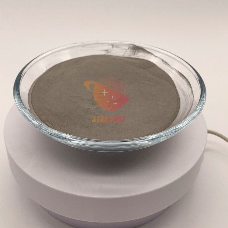
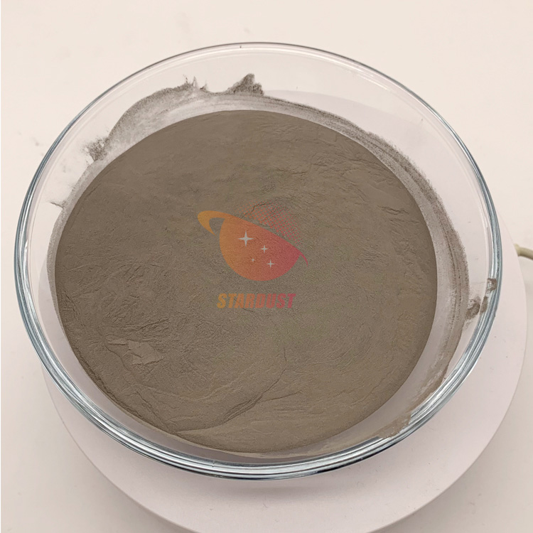
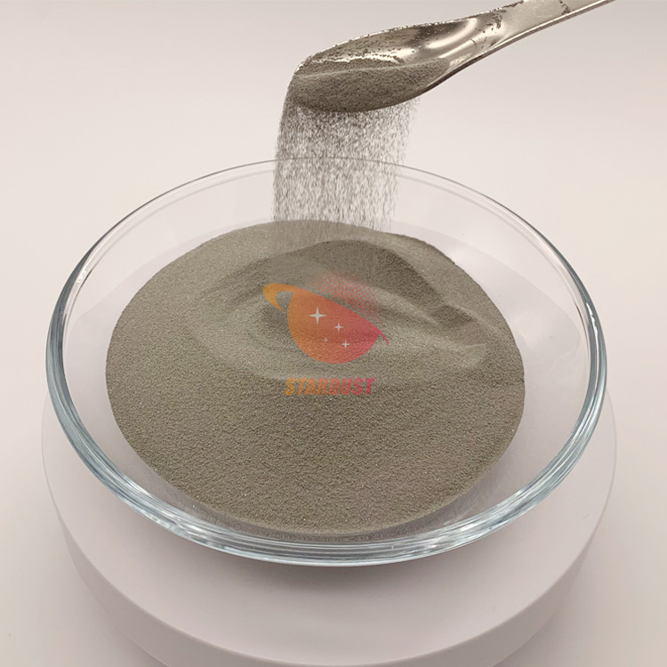
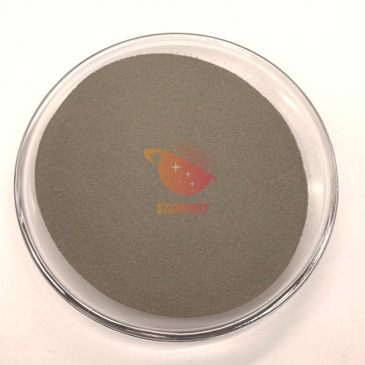
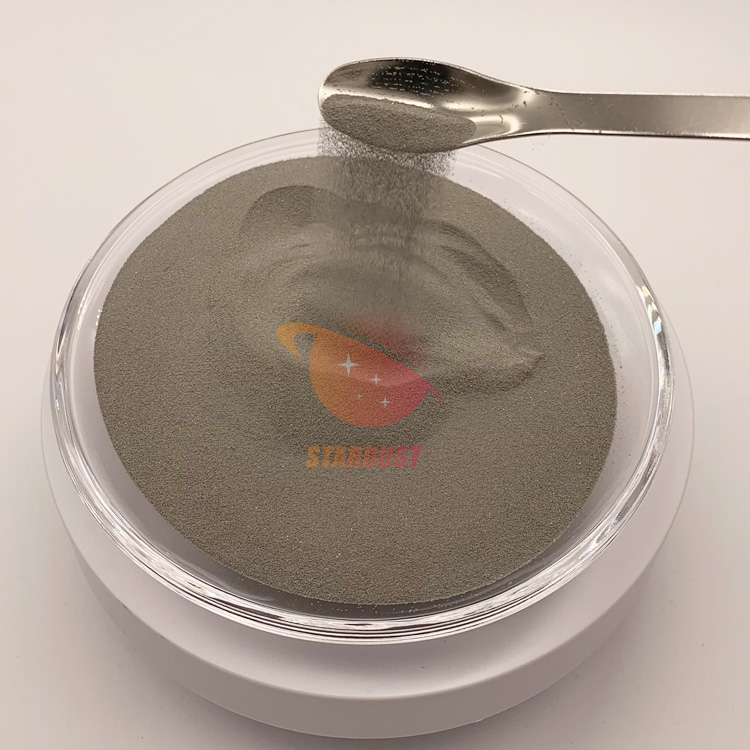
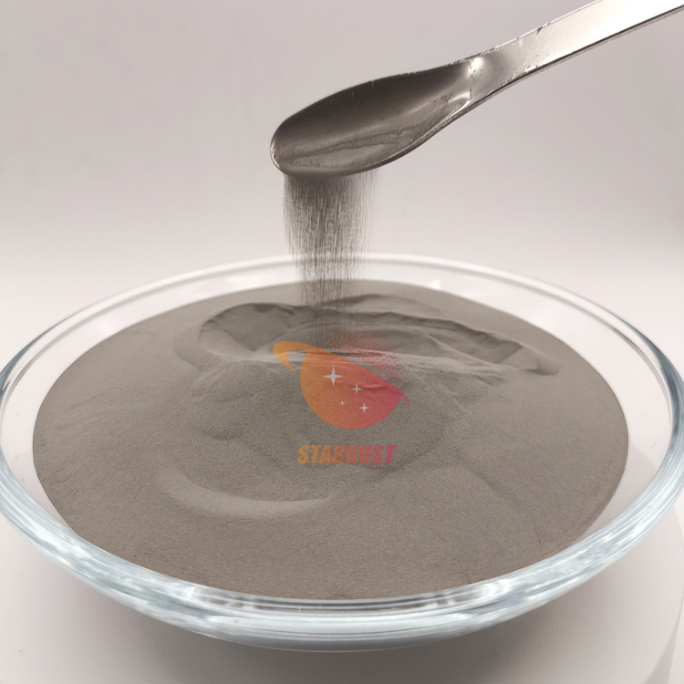
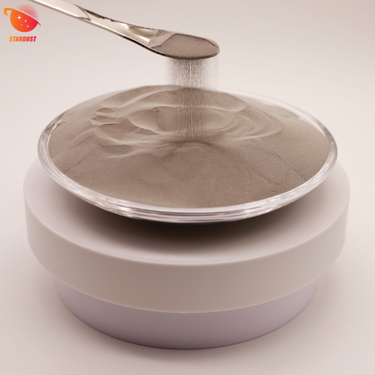
Stardust Technology's spherical tantalum powder is produced through radiofrequency plasma spheronization of high-purity tantalum. It exhibits outstanding powder characteristics: near-perfect spherical particles with uniform morphology and controllable particle size distribution; high bulk and tapped densities with excellent flowability, enabling uniform molding; Purity exceeds 99.95% with minimal impurities, ensuring outstanding chemical stability. It exhibits no leaching or oxidative corrosion in the human body environment. Additionally, it possesses excellent electrical conductivity, thermal conductivity, and mechanical plasticity. After sintering, the powder forms a porous structure with controllable porosity and pore size, meeting the requirements for medical forming processes.
In the field of medical implants, spherical tantalum powder is a core medical material, primarily used to prepare porous tantalum implants: sintered porous tantalum scaffolds, bone defect filling blocks, etc. Their pore structure matches human trabecular bone, facilitating bone cell adhesion, proliferation, and ingrowth to achieve osseointegration. This significantly enhances implant stability and reduces loosening risks. It is suitable for spacer repairs in hip and knee replacements, spinal fusion, and maxillofacial bone defect reconstruction. Additionally, high-purity spherical tantalum powder can be used to prepare medical coatings applied to implant substrates like titanium alloys, enhancing surface biocompatibility and corrosion resistance. Its outstanding biocompatibility also makes it suitable for dental implant components, meeting long-term human implantation requirements. Its biocompatibility and safety have been thoroughly validated in clinical applications. For more product information, please contact our specialist, Manager Cathie Zheng, at +86 13318326187.


 IPv6 network supported